Medical equipment status
- Positron emission tomography-CT (PET-CT)
- Magnetic resonance imaging (MRI)
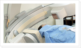
- Cardiovascular imaging device (ANGIOGRAPHY)
- Computed tomography (CT)
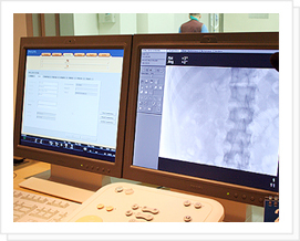
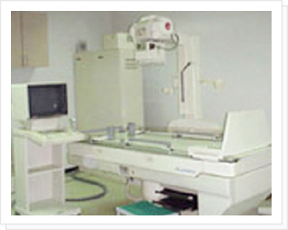
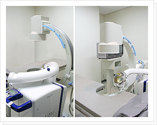
- X-ray for fluoroscopy (Remoter X-ray)
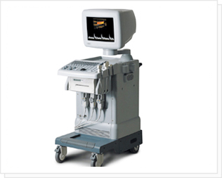
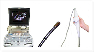
- Ultrasound diagnostic device (Ultra Sound)
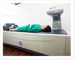
- Bone Densitometer
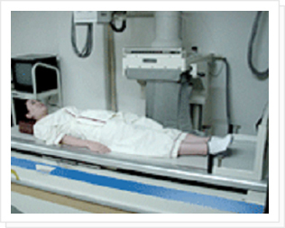
- Angiography X-ray
- extracorporeal shock wave lithotripter (Sonolith Model: Electroconductive Lithotripsy)
- Trans-esophageal Echocardiography (TEE)
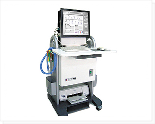
- Diabetic Cardiac Autonomic Neuropathy
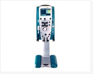
- CRRT: Continuous Renal Replacement Therapy
- Gamma Camera
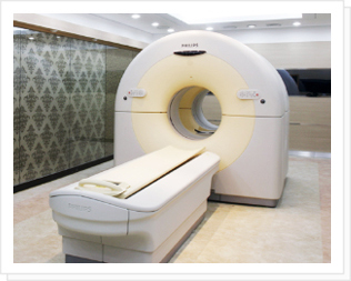
Positron emission tomography-CT (PET-CT)
- High-tech diagnostic imaging diagnostic equipment that combines the advantages of PET, which examines the abnormalities in the body’s metabolic activity, and CT, which examines the structural abnormalities in the body.
- With a single examination, it is possible to diagnose and detect cancer early, determine stages, and distinguish the rapid determination of metastasis, and it determines the location of cancer more accurately than conventional PET.
- Since the examination results of PET-CT can be directly used for medical treatment, the procedure for examination is simplified, and an efficient treatment plan can be established.
Diseases that can be diagnosed by PET-CT imaging
- Tumors (early diagnosis of cancer, distinguishment between benign and malignant tumors, recurrence evaluation, determination of treatment effect, etc.)
- Neuropsychiatric diseases (dementia, cerebrovascular disease, mental illness, cerebral palsy, epilepsy examination, etc.)
- Cardiovascular diseases (coronary artery disease, etc.)
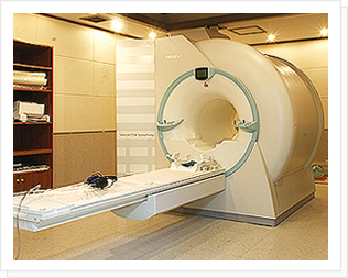
Magnetic resonance imaging (MRI)
- Using magnetic fields without harming the human body, it is possible to accurately diagnose the physical and chemical characteristics of human tissues with high-resolution 3D computer imaging. Patients can receive examinations on all parts of the body, including brain diseases, spinal diseases, musculoskeletal diseases, and organ diseases.
Related medical departments
Neurosurgery, Neurology, Orthopedics, Pediatrics, General Health Promotion Center, etc.
Cardiovascular imaging device (ANGIOGRAPHY)
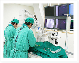
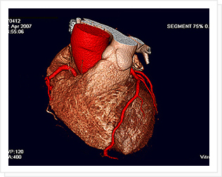
- This is the most accurate direct imaging method for diagnosing angina pectoris. Since the heart and blood vessels are beating continuously without a break, general X-ray examinations cannot properly diagnose and treat heart disease. Cardiovascular imaging device (angiography) is a high-tech medical equipment that can accurately diagnose and treat heart disease by taking dozens of digital images of the heart every second.
- In angiography using a cardiovascular imaging device, a long tube is inserted through a blood vessel in the groin or a blood vessel in the arm to the coronary artery, and a drug called a contrast medium is injected to directly photograph the blood vessel.
Related disease
heart and coronary artery disease
Related medical departments
Cardiology
Computed tomography (CT)
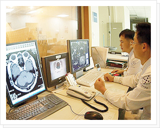
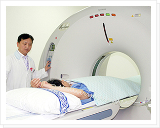
- Computed tomography (CT) refers to an imaging technique in which a target area of the human body is irradiated from various directions, the transmitted X-rays are collected with a detector, and the difference in X-ray absorption for that area is reconstructed by a computer using mathematical techniques. In addition, compared to conventional X-ray images, it has excellent resolution and contrast in distinguishing blood, cerebrospinal fluid, white matter, gray matter, and tumors, and can express minute absorption differences. Therefore, CT occupies a very important area in the field of imaging diagnosis.
- In particular, the CT possessed by our hospital is a 64-channel MDCT, which provides more accurate and clearer images than regular CT. In our hospital's radiology department, it is possible to obtain images that have the effect of endoscopy by implementing it as a 3D stereoscopic image.
Related medical departments
Neurosurgery, Neurology, Orthopedics, Pediatrics, General Health Promotion Center, etc.
X-ray for fluoroscopy (Remoter X-ray)
- It refers to transmitting X-rays generated by an X-ray generator through the human body and imaging the results on a film. It is used to observe the anatomical structure of the human body, the existence of bone fractures, the existence of abnormalities of soft tissues, and the progress of the affected area.
Ultrasound diagnostic device (Ultra Sound)
-
It is a device that transmits X-rays generated by an X-ray generator to the human body and images the results on a film. It is used to observe the anatomical structure of the human body, the existence of bone fractures, the abnormalities in soft tissues, and the progress of the affected part of the body.
Ultrasonic waves (a type of sound wave) that are harmless to the human body are projected into the body, reproducing the size and location information of the reflector caused by the difference in density between tissues as an image on the monitor.It is an essential examination of the internal medical area, including the liver, gallbladder, pancreas, spleen, and vascular system to diagnose the existence of diseases, in the urinary reproductive system to diagnose the size and shape of the lesions and distinguish abnormalities in the prostate and testicle, in gynecology including breast and pelvis, and in the field of obstetrics to detect abnormalities in the fetus.
Bone Densitometer
- Bone density refers to the hardness of the bone. The higher the bone density level means that the bone is harder. The purpose of measuring bone density is to prevent physical damage, economic loss, and shortening of life due to fractures by diagnosing and treating osteoporosis early.
Related disease
Osteoporosis
Related medical departments
General Health Promotion Center, Women’s Center, Endocrinology, Orthopedic Surgery, General Surgery, etc.
Bone density measurements must be taken for the following people:
- Women over the age of 65
- In case there is a risk of one or more osteoporotic fractures other than menopause
- Menopause before the age of 45
- Long-term use of steroids for arthritis
- Those whose mother had a femoral fracture or vertebral compression fracture
- Very thin person
- Patients with hyperthyroidism or hyperparathyroidism
- A person who spends a long time in bed laying down
- A person who spends a long time in bed laying down
- Short and stooped person
Angiography X-ray
- It is a blood vessel examination using radiation. A radiologist inserts a thin tube (about 2 mm) called a catheter from outside the body into the patient's blood vessel and injects a contrast medium. After that, the name of the disease, the location of the disease, and the progress of the disease are checked by determining whether the blood vessels are abnormal on the X-ray.
extracorporeal shock wave lithotripter (Sonolith Model: Electroconductive Lithotripsy)
- Using high-energy shockwaves, stone stones in the kidney, ureter, and bladder are turned into a fine powder and naturally excreted as urine. It is the safest and most effective high-tech medical device that does not require surgery or anesthesia.
Characteristics of treatment
- Hospitalization, skin incision, and anesthesia are not required.
- Daily life is possible immediately after 30 minutes of a procedure.
- Economical treatment method.
- The safest treatment method.
- No sequelae after the procedure.
- Safe treatment for the elderly and those with chronic diseases
- The sonolith vision model introduced in our hospital is a device made by introducing the most recent type of electroconductive lithotriptor (ECL) method. Compared to the conventional piezoelectric type, safety, and pain reduction effect are excellent. In particular, the ability to focus on stones and transfer energy, which can destroy stones accurately and effectively.
Related disease
Urinary stones (urethral stones, ureteral stones, bladder stones, kidney stones)
Related medical departments
Urology
Trans-esophageal Echocardiography (TEE)
- It is an examination that can obtain accurate information about the heart muscle, valves, heart function, and aorta located in the esophagus or upper stomach through an endoscopy.
- A limitation of transthoracic echocardiography is that it cannot be fully examined if the ultrasound beam does not pass well due to the patient's condition (e.g., the patient is obese or has chronic obstructive pulmonary disease). In this case, since the esophagus and the heart are attached, the heart can be observed very well without obstruction by placing an ultrasound machine in the esophagus. In addition, as the left atrium, which is farthest from the front chest, can be seen clearly, it is useful to find blood clots that can easily occur during atrial fibrillation.
People who:
- have chest pain and abnormal findings on the ECG
- with hypertrophy of the heart on chest X-ray
- have problems with the structure of the heart such as the atrium, ventricle, valves, and aorta
- with congenital heart disease or coronary artery disease
Related disease
Heart valve disease, myocardial infarction or weakness of the heart muscle, effusions of the pericardium and epicardium, blood clots in the heart, and other heart deformities
Related medical departments
Cardiology
Diabetic Cardiac Autonomic Neuropathy
- More accurate results can be obtained through the five existing examinations for early diagnosis of diabetic cardiovascular autonomic dysfunction and the newly added heart rate variability examination. Therefore, complications caused by diabetes can be prevented in advance. In addition, six examinations are performed at once, making it faster and more convenient.
People who have:
- Diabetic autonomic neuropathy and autonomic dysfunction
- Hypertension, orthostatic hypotension, and hypoglycemia
- Fatigue, depression, anxiety, and insomnia
- irritable bowel syndrome, and anorexia nervosa
Related medical departments
Endocrinology
“Diabetes is at risk of complications.”
CRRT: Continuous Renal Replacement Therapy
- CRRT is a treatment that purifies the blood in place of damaged kidneys by continuously circulating blood outside the body for 24 hours. It is a treatment method for acute renal failure accompanied by hemodynamic unstable multi-organ failure and various complications. It is mainly used in intensive care units.
Advantages of CRRT
- By reducing the burden on the cardiovascular system, it is possible to remove a large amount of water.
- It is easy to have electrolyte balance, acidity balance, volume control, etc.
- It is possible to remove inflammatory factors, has an excellent clearance rate, and has an excellent effect on preserving residual kidney function.
Gamma Camera
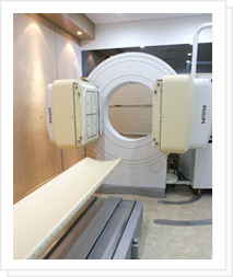
- Philips' Forte Dual Head Gamma Camera has been introduced and operated. Through SPECT inspection, high-resolution, three-dimensional images can be obtained in a shorter time. Nuclear medicine examinations are conducted on the thyroid, bone, heart, kidney, lung, brain, hepatobiliary tract, and liver as well as the digestive system, vascular system, tumor system, endocrine system, and immune system.


